Mostrar el registro sencillo del ítem
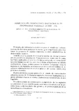
Observaciones microscópico-electrónicas en Trichomonas vaginalis donne 1.836
| dc.contributor.author | Bustos Ruiz, M. | es_ES |
| dc.date.accessioned | 2010-04-16T11:47:39Z | |
| dc.date.available | 2010-04-16T11:47:39Z | |
| dc.date.issued | 1976 | |
| dc.identifier.issn | 1885-4494 | |
| dc.identifier.uri | http://hdl.handle.net/10396/3014 | |
| dc.description.abstract | In the present research we have studied the fine structure of T. vaginalis isolated from patients with trichomoniasis. The protozoan was preserved by culture in vitro. The special reference is to the mastigont system, costa, parabasal body and the microtubular structure of axostyle and pelta. The undulanting membrane and flagella are also studied. | en |
| dc.description.abstract | Hemos estudiado la ultraestructura de T. vaginalis aislados de pacientes humanos afectados de tricomoniasis. Posteriormente se mantenían cultivos de este tricomonádido. Se hace principal referencia al sistema cinetosómico, la costa, el cuerpo parabasal y a la estructura micro tubular del axostílo y la pelta. También se describe la constitución de la membrana ondulante y de los flagelos, como principales orgánulos locomoción en este protozoo. | es_ES |
| dc.format.mimetype | application/pdf | es_ES |
| dc.language.iso | spa | es_ES |
| dc.publisher | Universidad de Córdoba, Servicio de Publicaciones | es_ES |
| dc.rights | https://creativecommons.org/licenses/by-nc-nd/4.0/ | es_ES |
| dc.source | Archivos de zootecnia 25 (100), 311-325 (1976) | es_ES |
| dc.subject | Microscopio electrónico | es_ES |
| dc.subject | Trichomonas Vaginalis | es_ES |
| dc.title | Observaciones microscópico-electrónicas en Trichomonas vaginalis donne 1.836 | es_ES |
| dc.title.alternative | Note on the electron-microscopical structure of Trichomonas vaginalis | en |
| dc.type | info:eu-repo/semantics/article | es_ES |
| dc.relation.publisherversion | http://www.uco.es/organiza/servicios/publica/az/az.htm | es_ES |
| dc.rights.accessRights | info:eu-repo/semantics/openAccess | es_ES |